Table of Contents
Introduction
Sonography, also known as ultrasound imaging, has long been an essential tool in modern medical diagnostics. It utilizes high-frequency sound waves to create real-time images of the body’s internal structures, allowing healthcare professionals to visualize and assess various organs and tissues. This non-invasive and radiation-free imaging technique has been instrumental in diagnosing a wide range of medical conditions, from monitoring fetal development during pregnancy to detecting diseases in various organs.
However, despite its crucial role in healthcare, traditional sonography has certain limitations and challenges. These include the reliance on human interpretation, the potential for variability in image quality, and the time-consuming nature of manually analyzing ultrasound images. This is where Artificial Intelligence (AI) has stepped in to bring about transformative breakthroughs in the field of sonography.

AI in Sonography
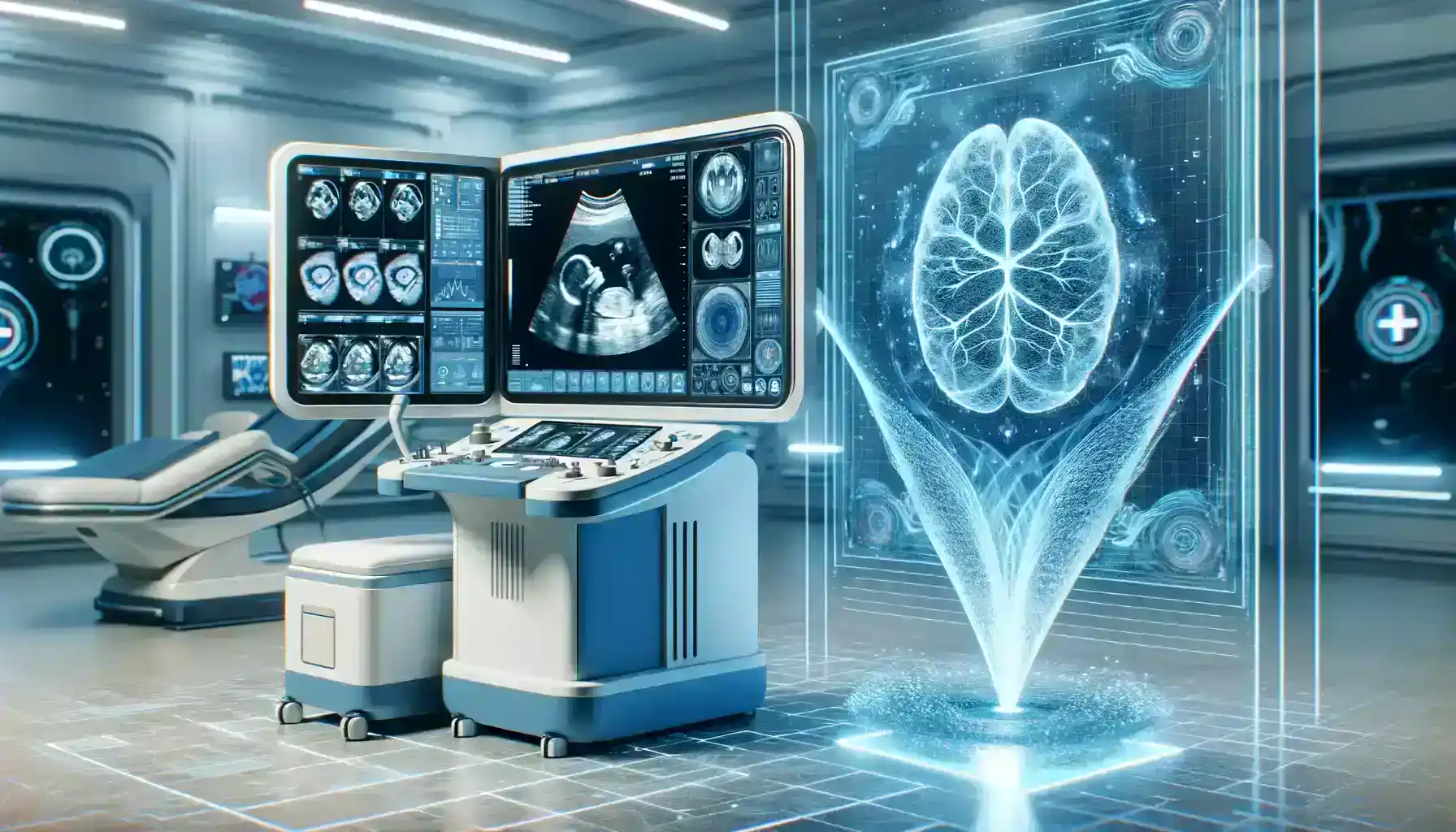
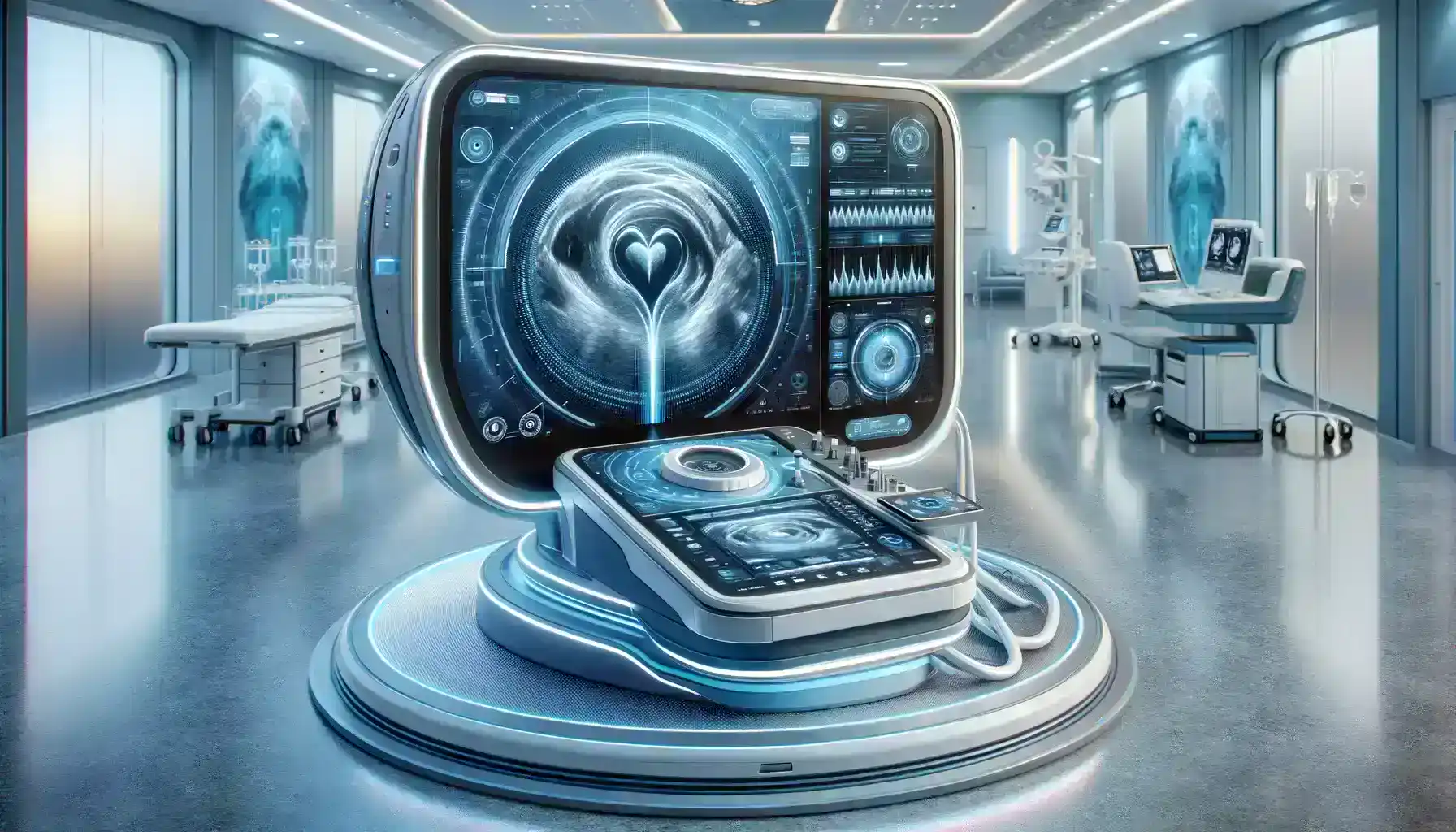
The integration of AI in sonography has brought about a series of significant advancements that are revolutionizing the way medical professionals use this imaging technique. AI in sonography is transforming the landscape of medical diagnostics. It’s not just a technological advancement; it’s a fundamental shift in how we leverage technology to improve patient care. As AI continues to evolve and be integrated into medical practice, it holds the promise of revolutionizing the field of sonography, ultimately benefiting patients and healthcare providers alike.
In this article, we’ll examine five key technological breakthroughs in the field of AI-assisted sonography.
1. Automated Image Analysis
EchoNous Vein AI is an AI tool designed to assist with vein assessment during ultrasound procedures. It helps locate veins and guides vascular access procedures.
Automated Image Analysis in Sonography is continually evolving and being integrated into ultrasound systems to enhance the accuracy and efficiency of diagnostic imaging. Healthcare providers and sonographers can explore these options to improve patient care and streamline the sonography process.
AI in sonography has revolutionized prenatal care by enhancing the accuracy of fetal anomaly detection, enabling earlier diagnoses, and ultimately improving patient care and outcomes. Automated image analysis powered by AI is transforming the way we approach healthcare, making it more efficient, reliable, and patient-centric.
2. Real-time Guidance
Cardiac sonography, also known as echocardiography, is a specialized field of sonography focused on imaging the heart. It plays a critical role in diagnosing heart conditions and guiding treatment decisions. The application of AI in cardiac sonography has introduced a transformative breakthrough, especially in real-time guidance.
Siemens Healthineers offers AI-powered solutions, including the AI-Rad Companion for Ultrasound Liver. This tool assists in liver imaging by providing real-time guidance for optimized image acquisition and quantification.
AI in Sonography, specifically real-time guidance, ensures that the cardiac ultrasound procedure is optimized for the best possible diagnostic outcomes. It assists the sonographer in obtaining precise views of the heart, leading to improved accuracy in diagnoses and more effective treatment planning. Patients benefit from faster and more accurate evaluations of their cardiac health, ultimately resulting in better healthcare outcomes.
3. Enhanced Image Enhancement
In vascular sonography, the visualization of blood flow in vessels is crucial for diagnosing and managing circulatory disorders. However, the quality of ultrasound images can be influenced by various factors, including the patient’s anatomy, the presence of obesity, or the depth of the targeted vessels.
Deep Vein Thrombosis (DVT) is a condition where blood clots form within deep veins, usually in the legs. Accurate and early diagnosis is essential to prevent potential complications like pulmonary embolism. AI in sonography can significantly aid in the detection of DVT by enhancing image quality.
The sonographer can then precisely identify the location and extent of the clot, facilitating prompt treatment. AI’s image enhancement not only improves diagnostic accuracy but also expedites the assessment process, leading to better patient outcomes.
4. Predictive Analytics for Disease Diagnosis
Breast cancer is a significant health concern for women worldwide, and early detection is crucial for improving treatment outcomes and increasing survival rates. Traditional breast cancer screening methods, such as mammography and ultrasound, have been effective but may sometimes result in false positives or false negatives. AI in sonography is changing the landscape of breast cancer detection by improving the accuracy and efficiency of breast ultrasound examinations.
By integrating AI-driven predictive analytics into breast ultrasound, healthcare providers can enhance their ability to detect breast cancer at its earliest stages. This technology not only improves diagnostic accuracy but also offers a more efficient workflow for healthcare professionals, ensuring that patients receive timely and appropriate care.
Butterfly iQ+ is an innovative handheld ultrasound device with integrated AI capabilities. It offers real-time guidance and automated measurements, assisting sonographers in capturing high-quality images and providing predictive analytics to aid in diagnosis.
5. Workflow Optimization
The integration of AI in sonography has not only enhanced the diagnostic capabilities of this medical imaging technique but has also significantly streamlined the workflow in healthcare settings. This breakthrough in workflow optimization is transforming how sonography is conducted and managed, with a particular focus on improving efficiency and patient care.
The incorporation of AI in sonography has not only advanced the diagnostic capabilities of this medical imaging technique but has also led to a profound transformation in workflow optimization. By automating routine tasks, reducing turnaround times, enhancing resource allocation, and minimizing patient wait times, AI-driven workflow optimization contributes to more efficient and patient-centered healthcare delivery.
Aidoc’s AI-powered radiology reporting platform automates the creation of radiology reports. AI-driven reporting software can automatically generate structured reports from imaging data, reducing the need for manual report creation.

Case Study
Transforming Neonatal Care: AI-Powered Sonography Saves a Tiny Life
In the world of neonatal care, every second counts. Premature infants often face critical health challenges, and rapid and accurate diagnosis is crucial for their survival. This case study showcases how AI-powered sonography played a pivotal role in saving a newborn’s life, demonstrating the profound impact of AI in sonography.
The Challenge: In a bustling neonatal intensive care unit (NICU), doctors and nurses were facing a recurring challenge: diagnosing and monitoring the health of premature infants. One particular case involved a baby girl, Emma, born prematurely at just 28 weeks gestation. Emma had difficulty breathing, and her oxygen saturation levels were dangerously low. The medical team suspected a cardiac condition, but conventional ultrasound imaging presented limitations in terms of speed and precision.
The Solution: The NICU introduced an AI-powered sonography system designed for rapid and accurate cardiac assessments in neonates. This innovative system incorporated real-time guidance, automated image analysis, and predictive analytics capabilities.
- Real-time Guidance: The AI system provided instant feedback to the sonographer during the cardiac ultrasound exam for Emma. It ensured that the transducer was positioned optimally to capture precise views of the heart, allowing for a faster and more accurate assessment.
- Automated Image Analysis: AI algorithms analyzed Emma’s cardiac images with unparalleled precision. They swiftly detected any abnormalities or structural issues within the heart, eliminating the potential for human error.
- Predictive Analytics: Machine learning models within the AI system assessed the data in real-time, comparing Emma’s heart structure to a vast database of neonatal cardiac conditions. This predictive analysis helped the medical team identify a rare congenital heart defect that had gone undetected in previous exams.
Results
- Swift Diagnosis: Emma’s congenital heart defect was diagnosed within minutes, allowing the medical team to initiate immediate treatment.
- Treatment Plan: Emma received specialized care tailored to her specific condition. Surgery was performed promptly to correct the defect, saving her life.
- Improved Outcomes: Thanks to the rapid and accurate diagnosis facilitated by AI, Emma’s health stabilized, and she eventually thrived, growing stronger with each passing day.
Emma’s case serves as a compelling method of the transformative power of AI in sonography. AI’s real-time guidance, automated image analysis, and predictive analytics capabilities proved to be life-saving tools in neonatal care.
The potential of AI’s Sonographic Marvel as a technological breakthrough holds the promise of revolutionizing neonatal care and countless other medical specialties. With AI’s assistance, healthcare professionals can provide more timely and accurate diagnoses, leading to improved patient outcomes and a brighter future for the world of medicine.

Conclusion
The synergy between Artificial Intelligence (AI) and sonography is nothing short of revolutionary. As demonstrated by Emma’s life-saving journey in neonatal care, AI in sonography stands as a marvel of modern medicine, a technological breakthrough that is redefining the possibilities of healthcare.
AI’s real-time guidance, automated image analysis, and predictive analytics have transformed the way we diagnose and treat patients, especially in critical situations like neonatal care. In Emma’s case, the rapid and precise diagnosis made possible by AI not only saved her life but also exemplified the tremendous potential of this technology to reshape the landscape of medicine.
The impact of AI in sonography extends far beyond Emma’s story. It promises improved patient care, enhanced diagnostic accuracy, and increased efficiency across various medical specialties. With each passing day, AI continues to push the boundaries of what is achievable in the field of sonography.
AI in sonography represents a remarkable leap forward in medical imaging, bringing us closer to a world where diagnoses are made swiftly, accurately, and with a profound impact on patient outcomes. The technological marvel that is AI in sonography is not just a breakthrough; it is a beacon of hope for the future of healthcare, where every second counts and every life matters.